

Road Founder Caused by Bad Trimming and Shoeing
|
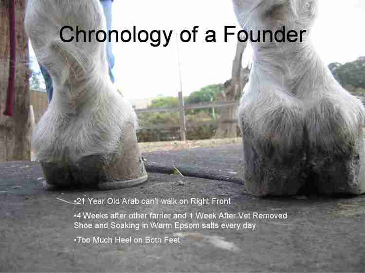
Another farrier shod the horse above 4 weeks before these pictures were taken. This horse was unable to bear any weight on the right front. The veterinarian removed the right front shoe a week before this picture was taken and requested that the owner soak the foot in warm Epsom salt water every day for 15 minutes and keep it in an Easy Boot. These pictures were taken before beginning work as a reference point for progress. |
|
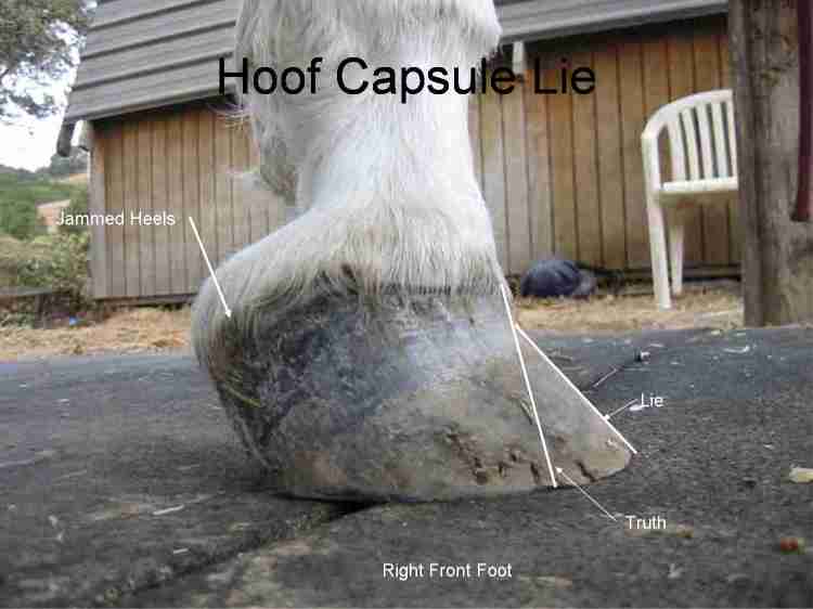
Both front heels are too high with evidence of severe jamming on the right foot along with possible delaminating of the hoof wall. The difference between the two lines drawn on the dorsal hoof wall suggest delamination (laminitis - separation of the pedal bone from the hoof wall) has occurred. |
|
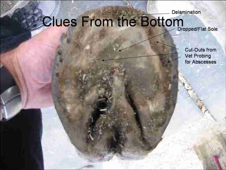
The sole is completely flat with a stretched white line (delamination). |
|
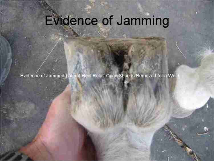
This picture above shows that the foot which has had its foot protected in an Easy Boot for a week has released toward the ground on the lateral (outside) of the foot once the shoe was removed. |
|
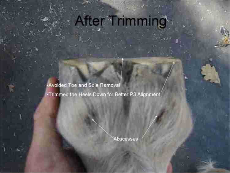
The heels were trimmed down, and this picture above shows the abscesses that the veterinarian was looking before on the sole of the foot. |
|
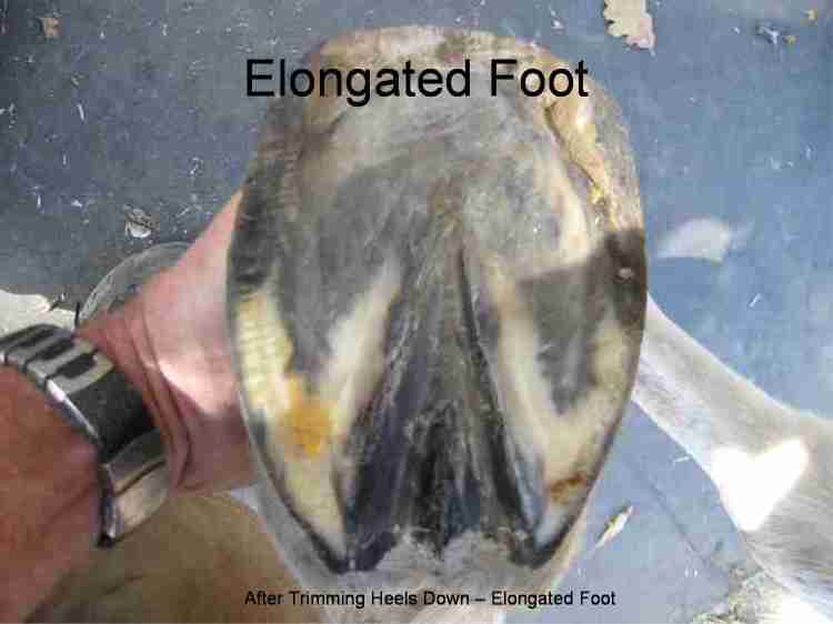
This is a view of the foot from the bottom after trimming. |
|
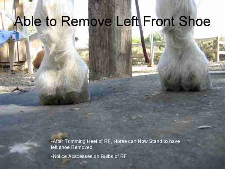
Before lowering the heel on the right front foot this horse could not stand on that foot to have the left front shoe removed. Trimming the heel down on this horse provided so much relief for this horse that he could then stand on the right foot to have the shoe removed on the left foot. |
|
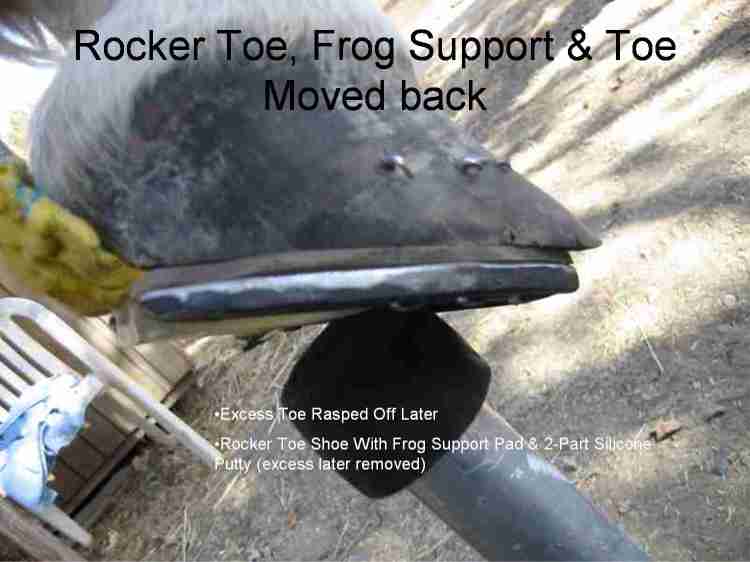
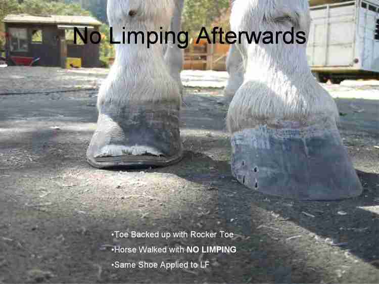
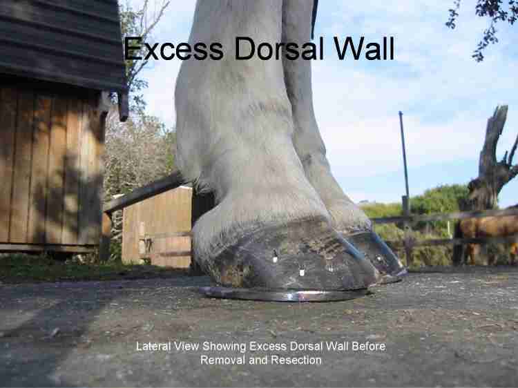
Even without radiographs, it was obvious that the break-over point needed to be moved back, so a rocker toe shoe with a frog support pad and two-part silicone putty was applied for support. The excess putty and dorsal wall were later removed as shown in the picture below. |
|
After moving the break-over point back and applying support for the frog (and the bone column), this horse walked normally without bobbing his head. |
|
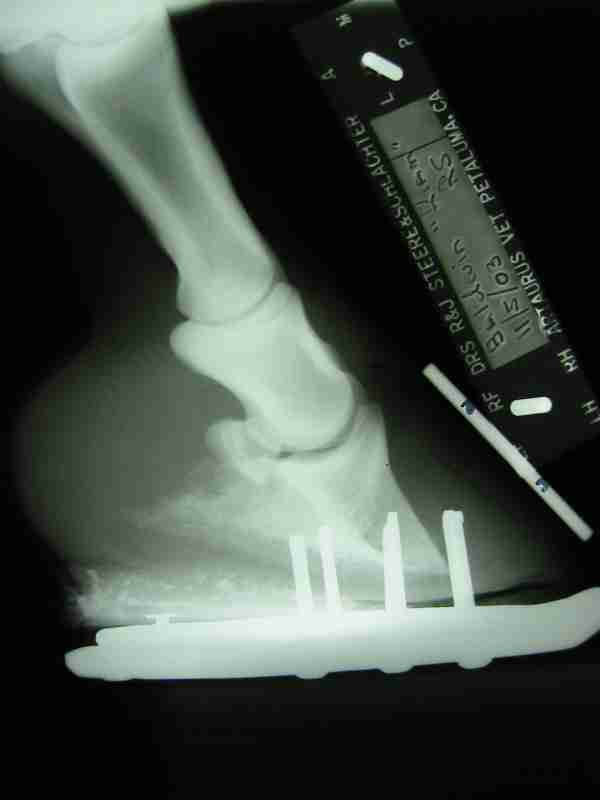
After 3 weeks the owner said that this gelding was completely sound at a walk but couldn't trot or run. The previous pictures were shared with the veterinarian, and radiographs were requested of the front feet which showed rotation and sinking of the coffin bone (aka pedal bone, P3, 3rd phalanx). I met in person with the following equine professionals at Farrier Focus in Fort Collins, Colorado to discuss the previous slides and the prognosis and direction to take for helping this gelding to survive this case of acute laminitis. Their opinions and advice are greatly appreciated.
|
|
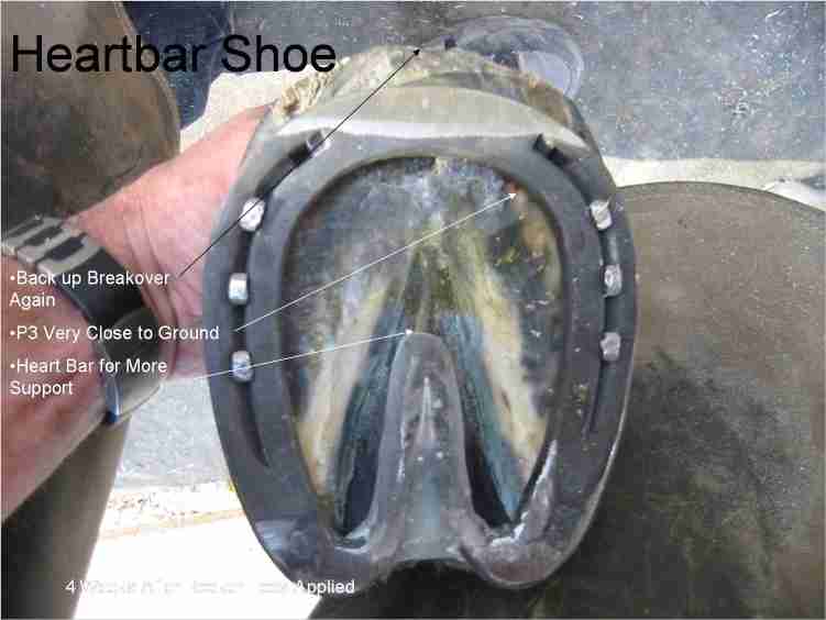
After sharing these pictures and radiographs with several equine professionals who specialize in laminitis, a decision was made to increase the support to P3 with a heartbar shoe and roller toes. The surface toward the sole was cupped to prevent any sole pressure. |
|
The shoes were again backed up to ease breakover and reduce pressure on the dorsal hoof wall. |
|
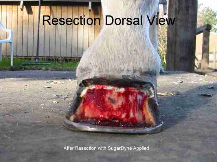
The dorsal hoof wall was resected with a dremel tool to allow the new hoof to grow out without any pressure from weight bearing that would cause further damage to the blood supply to the juvenile hoof wall at the coronary band. This gelding stood calmly and without pain as the hoof was removed. The red in the picture is not blood. It is sugardyne, a combination of betadyne and sugar used to prevent any infection. |
|
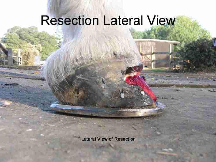
Notice how much the foot has been backed up at the dorsal wall compared to the previous pictures. |
|
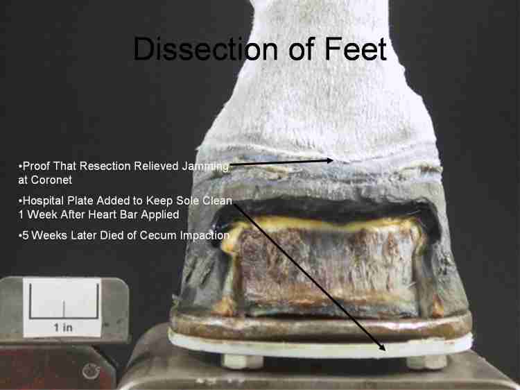
About 5 weeks later, this gelding died of a cecum impaction unrelated to the founder. The owner, who greatly loved this horse, made a decision to donate his front legs to be studied (dissected) so that other horses could benefit from what was learned about the condition that caused his laminitis. We took his front legs to Michael Savoldi, Chairman, of Equine Research Committee for the American Farriers Association, Shandon, CA, where he took several hundred pictures of this geldings feet as they were dissected. The mission of the Equine Research Committee is to encourage and develop research which specifically benefits the farrier. Notice in the picture above that the severe jamming at the coronary band at the front has begun to sag as a result of the resection--just as expected. The resection was successful. |
|
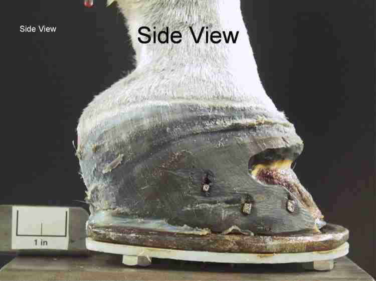
Notice the difference in the new growth rings in the hoof wall as they converge together at the toe. The blood supply was severely restricted by the jamming up of the hoof at the toe which suppressed growth in the dorsal (toe) area. |
|
Foot was completely sealed to reduce the possibility of infection from the ground to P3. |
|
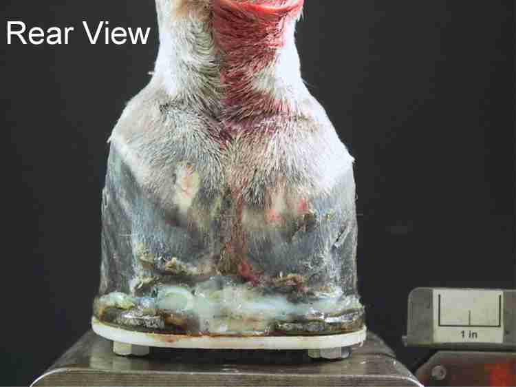
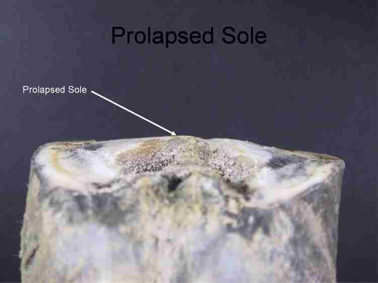
Evidence that the weight of the horse has been born by the sole. The sole should be cupped--not convex. |
|
In this picture (above) the heel is taken down to uniform sole thickness (UST), and the toe looks like it is rolling up in the air. Actually, the previous trim on this foot didn't left too much heel on this foot. Rasping at this angle caused a severe thinning of the sole in front of the frog and directly beneath the coffin bone which led to this gelding's lameness. The average sole is nearly 1/2 (.45") inch thick. When this sole was removed it was 1/16 inch to 1/8 inch thick--they were translucent at the point of P3 when held up to the light (after removing them). |
|
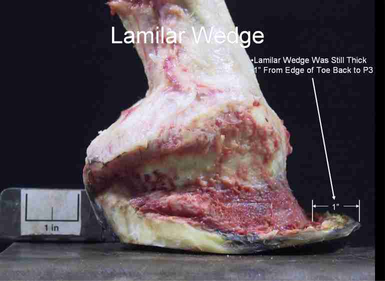
The lamina should be no more than 1/4" thick on this horse, but the distance from the edge of the lamilar wedge to the bone is at least 3/4" more than that. In other words, the resection shown in the previous pictures above were very safe to the horse and even more of the lamilar wedge could have been safely removed. |
|
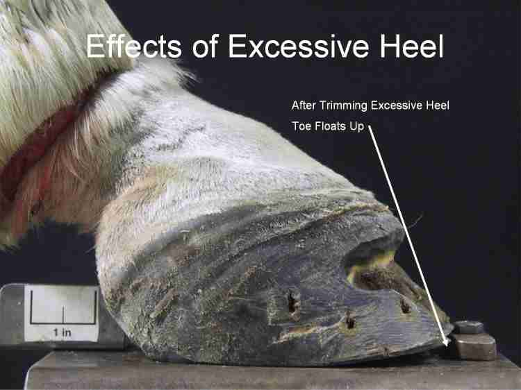
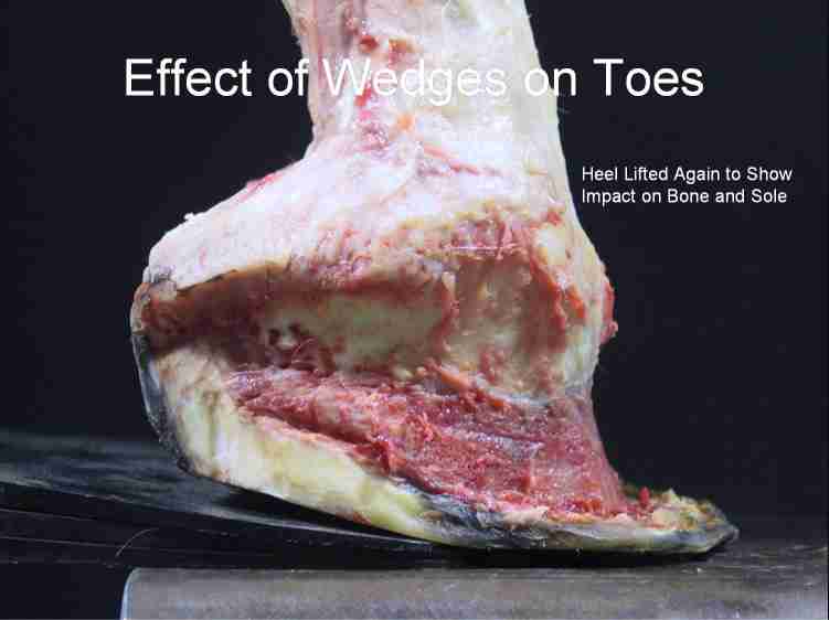
Look what wedging a heel does to the toe of a foot! It jams the bone into the sole over the solar corium and jams the dorsal hoof wall (toe) right up the front of the foot to the hair line. |
|
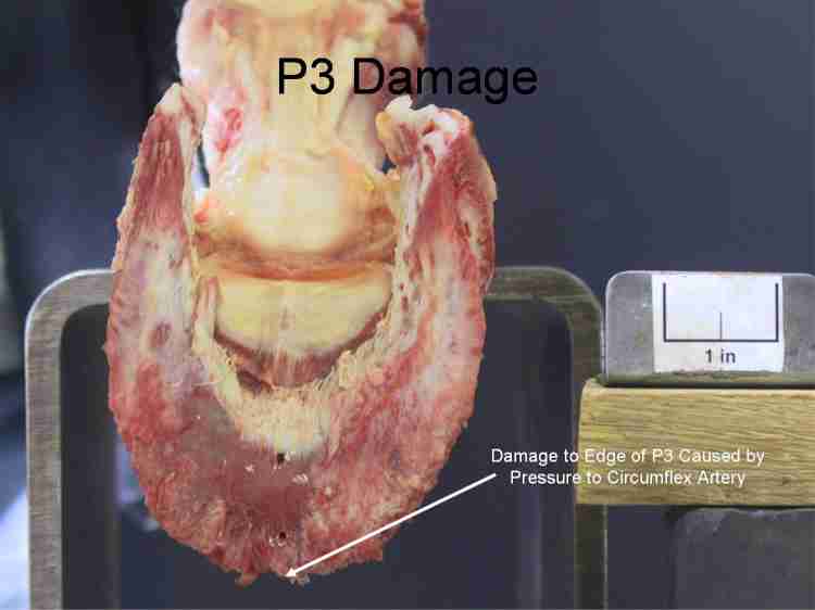
The rest of the hoof capsule was removed, and this picture shows the damage done to P3 at the tip of the bone. When P3 crushed the circumflex artery (which supplies blood to P3) on this horse, causing necrosis, the edge of the bone died (the rough edges), which also showed up as abscesses on the bulbs in the previous pictures. |